MRI - Magnetic Resonance Imaging
MRI (Magnetic Resonance Imaging) is one of the most sensitive diagnostic tools.
This medical miracle was first used on humans in 1971.
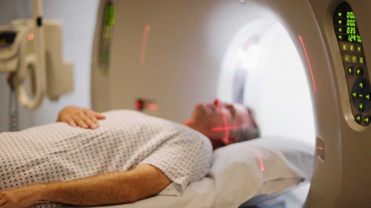
The final product reveals detailed anatomical images transferred onto film. MRIs differ from CT Scans in that there is no exposure to radiation. The MRI equipment is basically two powerful magnets; one external and one internal. Within the human body there are millions of negative and positive charged atoms. When these atoms are exposed to the electromagnetic waves produced by the MRI equipment, the atoms act like mini-magnets. By means of a computer, the data is collected, combined, and manipulated using complex mathematical equations. The final product reveals detailed anatomical images transferred onto film. MRI represents the gold standard in imaging. MRI is best for looking at soft tissues such as discs or nerves.
To appreciate the details rendered by an MRI consider the following contrast. Under x-ray, an intervertebral disc resembles a pocket of air. Using MRI the structure of the same disc is revealed in fine detail. Additionally, contrast dye introduced into the patient intravenously further defines and highlights particular aspects of the spine.
There are a few drawbacks to MRI. For example, take 100 normal people who appear to have nothing wrong with their spines and perform an MRI on each. The results may reveal that 20-25% of asymptomatic participants (without symptoms) have a herniated or bulging disc, or an arthritic condition. These patients are pain free and their lives go on without interruption at that particular time. The disadvantage is the results of an MRI may create a false positive. This means the MRI revealed a disorder for which there are no corresponding clinical symptoms. The point is this - the clinical symptoms must coincide with test results. It is not uncommon for a patient to come to the physician with a stack of MRIs indicating a herniated disc.
Lets say the patient is a competitive tennis player without clinical symptoms indicative of a herniated disc. In this case, to give the patient a serious diagnosis based simply on an MRI would be inappropriate. This is why MRI results must support the patient's clinical symptoms for a specific disorder. In some cases, a bulging disc does not cause any pain or problem. If leg pain is present and the MRI indicates a herniated disc associated with the nerves to the leg, it confirms the herniation as the cause of the leg pain.
For patients who are claustrophobic (claw-stro-foe-bick, fear of confinement) open-air MRI equipment is available. These patient-friendly imaging tables produce an excellent image without confinement in an imaging tube. Medicine to relax the patient is available and can be administered prior to the test.
Patients with internal ferromagnetic (metallic iron) devices such as a pacemaker, metal cardiac valve or metal in the area of the exam cannot be scanned. The powerful MRI magnets would interfere with these metal devices. In these patients a CT Scan is performed.

A National Practice
Dr. Eidelson sees patients from across the country for treatment of spinal stenosis and disc herniation. Most patients do NOT require surgery.
Call the Delray Beach office for information or to make appointment.