MRI for Patients
Magnetic resonance imaging (MRI) is type of diagnostic imaging. Unlike x-rays or computed tomography (CT or CAT) scans, an MRI does not use x-rays. It uses a powerful magnetic field and radio frequency pulses to produce very exact images of your internal organs, muscles, and bones. It is a valuable diagnostic tool.
MRI images can be printed out, displayed on a computer screen or be transmitted electronically from the radiologist to your physician. An MRI can produce very accurate images of your spine that can help your physician make a diagnosis or help plan for surgery. These images can be printed out, displayed on a computer screen, stored on a compact disk or flash drive, or be transmitted electronically from the radiologist to your physician. Magnetic resonance imaging produces vivid and complex images that can show your spine in great detail and allows your physician to see the relationship between your vertebrae, intervertebral discs, spinal cord, and nerve roots.
Where and How Is MRI Performed?
An MRI unit is large and is usually located in a hospital or a large radiology clinic. There are a few types of MRI. One type has a cylindrical tube that goes through a large circular magnet and you lie on a table that passes through the tube. A short-bore or open MRI unit uses a magnet that does not completely surround you, which can be helpful if you are larger than average or have claustrophobia. However, some types of MRI scans cannot be done with an open MRI unit.
If you are claustrophobic and know that you become nervous in small spaces, tell your physician. He or she may prescribe a tranquilizer to help you stay calm and relaxed during the scan.
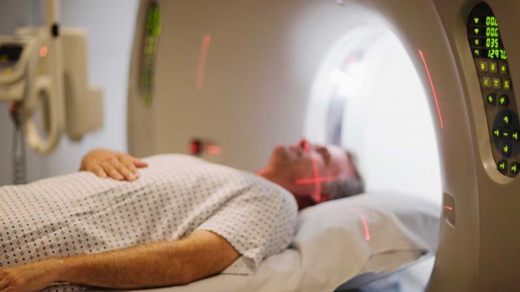
- You will lie on a table and the table will slide into the MRI unit.
- There may be specially shaped pillows or bolsters under you to help position you, depending on what kind of a scan is done.
As with a CT scan, the radiologist or technician may need to inject a contrast material, which is a dye that helps improve the visibility of certain parts of your spine. This will be injected through an intravenous (IV) line that will be inserted into your hand or arm before the scan starts. Often, your physician may want some MRI scans done before the contrast material is injected and after. After contrast medium is injected, you may feel a warm sensation or have a metallic taste in your mouth. If you have ever had a reaction to contrast medium in the past, make sure to tell your physician.
The technician will watch you throughout the exam from a control room. He or she can talk with you through an intercom. Periodically during the scan, you may be asked to hold very still or hold your breath for a few seconds.
While the scan is running, you will hear some moderately loud thumping or tapping noises. This is quite normal. You may be given earplugs or headphones to wear (listen to music).
Typically, an MRI takes up to an hour to complete. If your MRI includes contrast, the IV line is removed after the test.
How Should I Prepare?
You can usually drive to your MRI appointment. However, if you are claustrophobic and need a medication to help you relax during the procedure, you may be told to have someone drive you and bring you home again.
Before an MRI, you will change into a gown. You will be told to remove any metal objects such as jewelry, watches, hairpins, eyeglasses, and dentures. This is extremely important because you will be surrounded by a powerful magnetic field during the scan.
Important Considerations
If you have ever had an allergic reaction to any intravenous dyes or contrast material, you must tell your physician, the radiologist, or the technician.
It is very important that you tell your physician if you have any kind of metal implants in your body such as a pacemaker, dental implants, artificial joints, or aneurysm clips. Tell your physician if you have any metal shrapnel in your body. If you have any pieces of metal in your body, you probably cannot undergo magnetic resonance imaging.
What Happens Next?
A radiologist will look at the scans and analyze the images. He or she will send a report to your physician who will discuss the findings with you. You may be asked to have the MRI redone to get another look at something on your spine, to check for any changes, or to see how well a treatment is working.

A National Practice
Dr. Eidelson sees patients from across the country for treatment of spinal stenosis and disc herniation. Most patients do NOT require surgery.
Call the Delray Beach office for information or to make appointment.